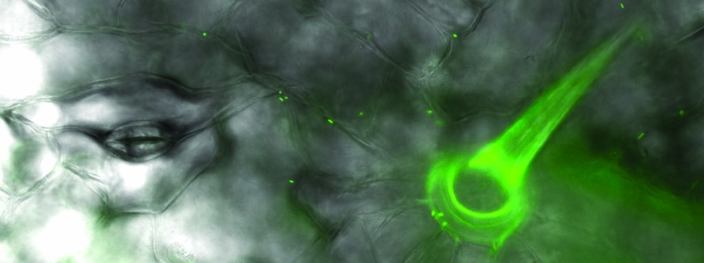
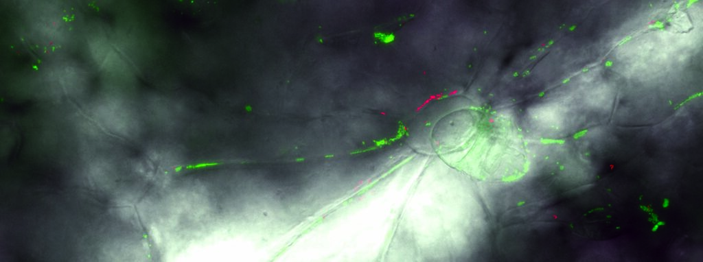
Images prises au microscope en épifluorescence. Bactéries de la phyllosphère Pantoea agglomerans (exprimant la protéine de fluorescence verte) et Pseudomonas syringae (exprimant la protéine de fluorescence rouge) sur la surface d’une feuille de haricot vert. Les structures végétales visibles sont les cellules de l’épiderme, des stomates, et des trichomes.
Phyllosphere bacteria
Images taken with epifluorescence microscopy. Phyllosphere bacteria Pantoea agglomerans (expressing the green fluorescent protein) and Pseudomonas syringae (expressing the red fluorescent protein) on the surface of a green bean leaf. Visible plant structures are the epidermal cells, stomates, and trichomes.
Of Bacteria and Men: Modelling the bacterial colonization of leaves


